Introduction
Kidney biopsy remains a cornerstone diagnostic tool in pediatric nephrology [1], providing crucial insights into the underlying pathology of various kidney diseases. While non-invasive diagnostic methods have evolved, histopathological examination continues to guide therapeutic decisions and prognostication in children with kidney disorders. Additionally, the advent of electron microscopy has revolutionized our understanding of podocytopathies and their variants. National registries of kidney biopsies showed a variety of kidney diseases and different epidemiology worldwide [2, 3, 4, 5, 6]. This single-center study aims to analyze the clinicopathological spectrum of pediatric kidney diseases based on native kidney biopsies, emphasizing the correlation between clinical presentations and histopathological findings. The study highlights the need of routine electron microscopy in pediatric renal biopsies. Hence, we aimed to characterize the indications and renal histopathological spectrum of kidney diseases among children who underwent kidney biopsy in Sheri-Kashmir Institute of Medical Sciences, a tertiary Care Center in North India.
Subjects and methods
This retrospective single-center, cross-sectional study was conducted at the Department of Nephrology in a tertiary care center in North India. The data of the children aged ≤18 years who underwent kidney biopsy in the Department of Nephrology from January 2022 to August 2024 was retrieved from the hospital records and analyzed. The study is approved by the institutional ethics committee of Sheri-Kashmir Institute of Medical Sciences under Reference no. SIMS/131/IEC-SKIMS/2024-209 dated 03 September 2024.
Participants
We collected data of all those who underwent renal biopsies from January 2022 to August 2024. The collected data included the age at presentation, symptoms, clinical picture, lab investigations, response to treatment, and indication for kidney biopsy. The indications of renal biopsy included nephrotic syndrome (NS) with atypical features like age <1 year or >12 years, steroid resistant nephrotic syndrome, nephrotic syndrome with renal dysfunction, nephrotic syndrome with systemic features, nephrotic syndrome with hypertension, nephritic syndrome, asymptomatic urinary abnormalities, acute kidney injury (AKI), chronic kidney disease (CKD) with normal size kidneys, and nephrotic-nephritic syndrome (nephrotic syndrome with active urinary sediment). Nephrotic syndrome was defined as urinary protein excretion of >40 mg/m2 in a 24-hour urine collection, or spot protein creatinine excretion ratio of >2000 mg/g with generalized edema and hypoalbuminemia. Steroid-resistant NS was defined as persistent proteinuria of more than 40 mg/m2 per hour after 4 weeks of prednisone therapy. Nephritic syndrome was defined as the presence of new-onset edema, hypertension, hematuria, and sub-nephrotic proteinuria. Asymptomatic urinary abnormality is defined as the presence of abnormal urine sediment in the form of proteinuria and/or microhematuria in the absence of any obvious clinical symptom or noticeable clinical signs.
Methods
As per the Department policy, all Kidney biopsy samples are sent for light microscopy, immunofluorescence microscopy, and electron microscopy. Immunological evaluation in the form of antinuclear antibody (ANA), complement protein 3 and 4 (C3, C4), anti-myeloperoxidase antibody (Anti MPO), anti-proteinase-3 antibody (Anti PR3), anti-glomerular basement membrane antibody (Anti GBM), and anti-streptolysin O antibody (ASO) titers is done based on the clinical scenario. Kidney biopsy is done under sedation with ketamine at a dose of 1–3 mg/kg body weight using an automated spring-loaded Bard max-core disposable biopsy instrument sized 16 × 18 G manufactured by Bard Peripheral Vascular, a US-based company. All biopsies are done under real-time ultrasound guidance using ESAOTE SpA (Ultrasound machine), an Italian company located at Via E. Melen, 77, 16152 Genova, Italy. Transmission electron microscopy is done using a 120 kV JEOL Transmission Electron Microscope (TEM) (TEM, Akishima, Tokyo). Tissue for light microscopy is fixed using 10% neutral buffered formalin. Serial sections of 2–3 μm thickness are made with 8 to 16 paraffin sections per biopsy, and at least 2 sections are placed per slide. Staining is done using Hematoxylin and Eosin (H&E), Periodic acid Schiff (PAS), Silver Methenamine, Masson Trichrome (MT), and Congo Red. Tissue for immunofluorescence is sent in Michels transport medium [7]. The tissue is frozen and cut into 2–4 μm sections in a cryostat. Immunostaining is done using antisera for IgA, IgG, IgM, C3, C4, C1q, and Kappa and Lambda light chains. Tissue for Electron microscopy (EM) is fixed in 2% glutaraldehyde. Tissue is cut into slices measuring 1–2 mm in thickness. The tissue is dehydrated and embedded in plastic resin, and semi-thin and thin sections are made, and the sections are placed on copper grids and stained with uranyl acetate and lead citrate. One glomerulus and accompanying tubule-interstitium is evaluated. The immunohistochemical markers like Vimentin, CK7 (cytokeratin 7), Carbonic anhydrase IX, Kidney-specific cadherin, and CD117 (cluster of differentiation 117) were not included. Patients with biopsy-proven lupus nephritis were classified based on the International Society of Nephrology and Renal Pathology Society classification (ISN/RPS) [8].
Statistical methods
The numerical data are presented as mean ± SD (standard deviation). Categorical variables are presented as percentages. The Mann-Whitney U test is used for non-parametric numerical data. Statistics are done using IBM SPSS (Statistical Package for the Social Sciences) version 25, developed and marketed by IBM Corporation, based in Armonk, NY, USA.
Results
Out of 601 biopsies done during the study period, pediatric biopsies accounted for 61/601 (10.1%) of all biopsies. Baseline characteristics are shown in Table 1.
Table 1: Baseline characteristics at the time of biopsy.
| Parameter | Value | |
| Total Biopsies | 601 | |
| Pediatric Biopsies | 61 (10.1%) | |
| Boys | 29 (47.5%) | |
| Girls | 32 (52.5%) | |
| Mean Age in years | 12.1 ± 5.1 | |
| Proteinuria (mean ± SD) grams per day | 4.4 ± 3.7 | |
| Creatinine (mean ± SD) mg/dL | 1.14 ± 1.6 | |
| Urea (mean ± SD) mg/dL | 31.9 ± 28 | |
| Hypertension | 23 (37.8%) | |
| Hematuria | 26 (42.6%) | |
| Headache | 6 (9.8%) | |
| Age Group (yr) | ||
| ≤5 | 10 (16.4%) | |
| 5–12 | 17 (27.9%) | |
| 12–18 | 34 (55.7%) | |
SD: Standard deviation.
The most common indication for kidney biopsy was nephrotic syndrome, present in 39/61 (63.9%), followed by nephritic syndrome and asymptomatic urinary abnormality (Table 2).
Table 2: Clinical indications for kidney biopsy.
| Indication of biopsy | Number of Cases | Percentage (%) |
| Nephrotic Syndrome | 39 | 63.9% |
| Nephritic Syndrome | 9 | 14.8% |
| Asymptomatic Urinary Abnormality | 5 | 8.2% |
| AKI | 3 | 4.9% |
| CKD | 3 | 4.9% |
| Nephrotic-nephritic syndrome | 2 | 3.3% |
| Total | 61 | 100.0% |
AKI: Acute kidney injury; CKD: Chronic kidney disease.
The most common indication for kidney biopsy in the nephrotic sub-group was onset beyond 12 years of age, followed by active urinary sediment (Table 3).
Table 3: Indications for kidney biopsy in Nephrotic syndrome.
| Characteristic | Frequency | Percentage |
| Age <1 yr | 1 | 2.6% |
| Age >12 yr | 18 | 46.1% |
| Active urinary sediment | 12 | 30.8% |
| Hypertension | 9 | 23.1% |
| SRNS | 8 | 20.5% |
| Systemic features | 2 | 5.1% |
Patients had ≥1 indication for biopsy. SRNS: Steroid-resistant nephrotic syndrome.
The most common overall histopathological pattern of injury was minimal change disease, present in 18/61 (29.5%), followed by FSGS and IgA nephropathy (Table 4).
Table 4: Histopathological pattern of injury.
| Histopathological Diagnosis | Number of Cases (%) |
| Minimal Change Disease (MCD) | 18 (29.50) |
| Focal Segmental Glomerulosclerosis (FSGS) | 12 (19.70) |
| IgA Nephropathy | 8 (13.10) |
| Lupus Nephritis | 5 (8.20) |
| Acute tubular injury | 3 (4.90) |
| MPGN | 2 (3.27) |
| IRGN | 2 (3.27) |
| C1q Nephropathy | 2 (3.27) |
| MN | 1 (1.64) |
| DN | 1 (1.64) |
| Global glomerulosclerosis | 1 (1.64) |
| Pauci-immune Glomerulonephritis | 1 (1.64) |
| CIN | 1 (1.64) |
| Fabry’s disease | 1 (1.64) |
| Unclassified collagenopathy | 1 (1.64) |
| Alport’s | 1 (1.64) |
| Morphologically normal | 1 (1.64) |
| Total | 61 (100.00) |
MN: membranous nephropathy; DN: Diabetic nephropathy; MPGN: Membranoproliferative glomerulonephritis; IRGN: Infection-related glomerulonephritis; CIN: Chronic interstitial nephritis; IgA: Immunoglobin A.
Minimal change disease, FSGS, and IgA nephropathy accounted for 18/39 (46.2%), 10/39 (25.6%), and 5/39 (12.8%) of all cases of nephrotic syndrome (Table 5).
Table 5: Clinical syndrome and histopathological diagnosis.
| Clinical Syndrome | Histopathological Diagnosis | Total | Percentage |
| Nephrotic syndrome | |||
| Minimal Change Disease (MCD) | 18 | 46.2% | |
| Focal Segmental Glomerulosclerosis (FSGS) | 10 | 25.6% | |
| IgA Nephropathy | 5 | 12.8% | |
| C1q Nephropathy | 2 | 5.1% | |
| MN, MPGN, Alport’s syndrome, & Global sclerosis | 1 each | 39/61 | |
| Nephritic Syndrome | |||
| Lupus Nephritis | 3 | 9/61 | |
| IgA Nephropathy | 3 | ||
| IRGN | 1 | ||
| MPGN | 1 | ||
| ANCA Vasculitis | 1 | ||
| Asymptomatic urinary abnormality | |||
| FSGS | 2 | 5/61 | |
| Collagenopathy | 1 | ||
| Lupus Nephritis | 1 | ||
| Normal histology | 1 | ||
| AKI | |||
| Acute tubular injury | 3 | 3/61 | |
| CKD | |||
| CIN | 1 | 3/61 | |
| Fabrys disease | 1 | ||
| Diabetic Nephropathy | 1 | ||
| Nephrotic-Nephritic | |||
| IRGN | 1 | 2/61 | |
| Lupus Nephritis | 1 | ||
AKI: Acute kidney injury; ANCA: Anti-neutrophil cytoplasmic antibody; CIN: Chronic interstitial nephritis; CKD: Chronic kidney disease; IRGN: Infection-related glomerulonephritis; MN: Membranous Nephropathy; MPGN: Membranoproliferative glomerulonephritis; IgA: Immunoglobin A. All kidney biopsies were assessed by Light microscopy, Immunofluorescence, and Electron microscopy. Staining was done using Hematoxylin and Eosin (H&E), Periodic acid Schiff (PAS), Silver Methenamine, Masson Trichrome (MT), and Congo Red.
Out of 9 patients with nephritic syndrome, IgA and lupus nephritis each accounted for 3/9 (33.3%), followed by infection-related glomerulonephritis, immune complex Membranoproliferative glomerulonephritis (MPGN) pattern of injury, and Anti-neutrophil cytoplasmic antibody (ANCA)-associated vasculitis, each contributing 1/9 (11.1%). The clinical and demographic profile of Minimal Change Disease (MCD) and FSGS is shown in Table 6. All MCD cases demonstrated diffuse foot process effacement on electron microscopy, minimal interstitial fibrosis/tubular atrophy (<10%), and presented with nephrotic syndrome. Table 6 below.
Table 6: Comparison between Minimal Change Disease (MCD), and Focal Segmental Glomerulosclerosis (FSGS).
| Characteristic | Minimal Change Disease (MCD) | Focal Segmental Glomerulosclerosis (FSGS) | p-value | |
| Prevalence in Biopsies | 18/61 (29.5%) | 12/61 (19.7%) | ||
| Gender Distribution | ||||
| Boys | 11 (61.1%) | 8 (66.7%) | 0.534 | |
| Girls | 7 (38.9%) | 4 (33.3%) | ||
| Age Distribution | ||||
| ≤5 yr | 7/18 (38.9%) | 3/12 (25.0%) | 0.626 | |
| 5–12 yr | 5/18 (27.8%) | 3/12 (25.0%) | ||
| 12–18 yr | 6/18 (33.3%) | 6/12 (50.0%) | ||
| Common Presentation | Nephrotic syndrome in all cases (100%) | Nephrotic syndrome in 10/12 (83.3%) | 0.152 | |
| Histopathological Findings | Diffuse foot process effacement on electron microscopy; minimal interstitial fibrosis/tubular atrophy (<10%) | Diffuse foot process effacement in 8/12 (66.7%) | ||
| Mean Age | 10.3 ± 5.3 yr | 11.3 ± 6.1 yr | 0.188 | |
| Median Serum Creatinine | 0.37 mg/dL | 0.45 mg/dL | 0.242 | |
| Mean Proteinuria | 5.6 ± 3.9 g | 4.6 ± 2.1 g | 0.966 | |
| Variants | Not specified | NOS variant: 10/12 (83.3%); tip and perihilar variants each represented one case | ||
| Clinical Significance | Commonly associated with nephrotic syndrome | More common in adolescents; and may indicate a more severe renal disease | ||
NOS: Not otherwise specified.
Chronic kidney disease secondary to diabetic nephropathy (resulting from type 1 diabetes mellitus), Fabry’s disease, and chronic interstitial nephropathy (CIN) was identified in three patients. The mean age and proteinuria in the CKD group were 16.3 ± 1.16 years and 1.39 ± 0.70 gram respectively. The median creatinine was 2.7 mg/dL. IgA nephropathy demonstrated male predominance (5/8; 62.5%) with a nephrotic presentation in 5/8 (62.5%) of cases. Gross painless hematuria was the presenting complaint in 2/8 (25%) patients with IgA nephropathy. Mean age at the time of biopsy was 12 ± 3.5 years. Among those with the nephrotic presentation, 60% showed diffuse foot process effacement, suggesting a podocytopathic variant of IgA nephropathy (IgAN). Lupus Nephritis exclusively affected female adolescents (12–18 years), with nephritic syndrome being the primary presentation (60%). The ISN-RPS classification revealed Class IV in two cases, Class II in one case, Class III in one case, and Class IV + V in one case. Out of 61 patients, 2 patients (3.3%) developed gross hematuria, and 5 patients (8.2%) developed perinephric hematoma.
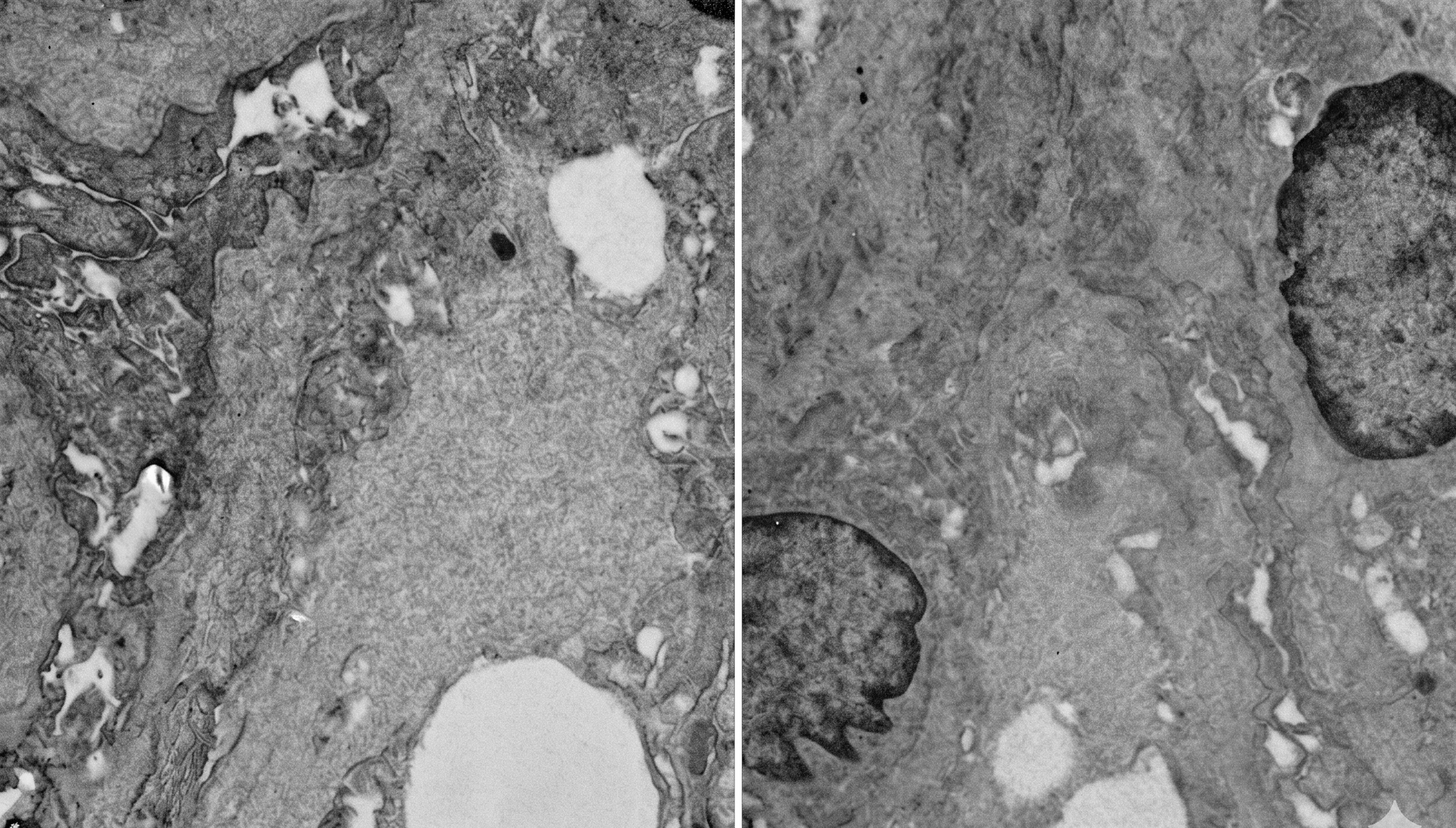
The patient with Fabry’s disease was a 15-year-old male who presented with bilateral lower limb swelling and newly detected hypertension. Evaluation revealed sub-nephrotic proteinuria (1 gram/day) with active urinary sediment and renal dysfunction with creatinine of 2.70 mg/dL. Kidney biopsy showed an enlarged and fine vacuolated appearance of podocytes, suggestive of Fabry’s disease. The patient with Alport’s disease was a 13-year-old male who presented with bilateral lower limb swelling, and evaluation revealed nephrotic syndrome (24 h urine protein 8 grams/day). He underwent a biopsy in view of failure to achieve remission after 4 weeks of steroid therapy (SRNS). The kidney biopsy showed a FSGS pattern of injury on light microscopy, and Electron microscopy showed glomerular basement membrane wrinkling and basket weave appearance suggestive of Alport’s syndrome. The patient was subsequently found to have anterior lenticonus and bilateral sensorineural hearing loss. Figs. 1,2 show photomicrographs from a patient with Fabry’s disease and Alport’s syndrome respectively.
Figure 1 PAS-Stained glomerular sections showing foam cell storage pathology. Left panel shows prominent clear vacuolated foam cell accumulation within glomerular capillary loops. Right panel shows extensively involved glomerulus with diffuse foam-cell accumulation and expansion of the glomerular tuft.
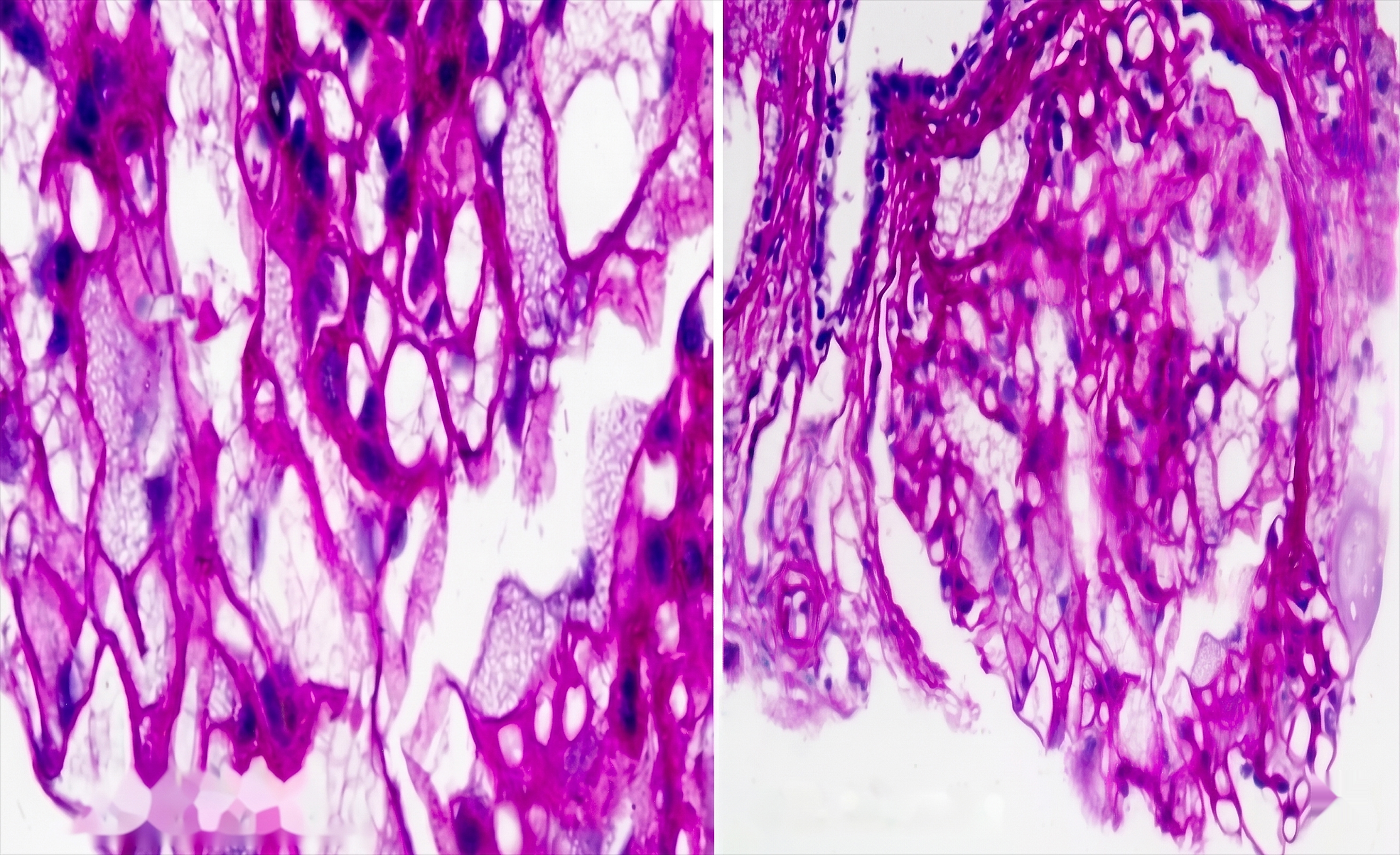
Figure 2 Transmission Electron Micrographs showing ultrastructural glomerular basement membrane abnormalities consistent with Alport’s syndrome. Left panel shows Irregular GBM wrinkling and lamellation. Right panel shows diffuse GBM multilamellation with a basket-weave appearance.
Discussion
Our study demonstrates that pediatric cases constituted 10.1% of all kidney biopsies performed at our center. This percentage is higher than the 4% reported by Yadav Subhash et al. [9] from Mumbai, India, who analyzed biopsy data collected over 6 years. This might be due to regional variations in biopsy practice or due to true differences in the incidence of pediatric kidney disorders based on environmental and genetic factors. The slight female predominance (53.5%) in our cohort differs from some earlier studies showing male preponderance [10, 11] and coincides with a few studies showing female preponderance [12, 13]. The female preponderance in our study is likely a chance finding. The mean age in our study is 12.1 ± 5.1 years, which is similar to the mean age of 11 ± 5 years reported by Alhasan K [14]. This may be attributed to our biopsy protocol, wherein all children with nephrotic syndrome aged above 12 years were biopsied, while those aged 1 to 12 years with steroid-sensitive nephrotic syndrome were not biopsied. This selective approach likely indicates a potential selection bias in the study population. Nephrotic syndrome emerged as the predominant indication for biopsy (63.9%). In the majority of the published reports about kidney biopsy in children, NS was the most common indication for biopsy, accounting for 28.5%–74.2% of patients, consistent with our results [10, 15, 16, 17]. The kidney biopsies were done for diagnostic purposes in all the cases. The most common reason for biopsy in children with nephrotic syndrome in our cohort was age beyond 12 years in 29.5%, followed by active urinary sediment, and associated hypertension in 19.6% and 14.8%, respectively. SRNS was the indication in 13.1% children. At our center, a kidney biopsy is done in nephrotic syndrome with atypical features like the presence of hypertension, renal dysfunction, active urinary sediment, associated systemic manifestations, steroid resistance, and steroid dependence, and if onset is beyond 12 years of age.
The knowledge of histopathological diagnosis is critically important from a clinical standpoint. The decision about further evaluation, the choice of treatment, and prognosis depends upon the histopathological pattern of injury. The patients with nephrotic syndrome secondary to Alport’s syndrome and Fabry’s disease were continued on Ramipril 5 mg daily, and no further immunosuppression was given. Immunosuppression was not given to patients with FSGS lesions and focal foot process effacement on electron microscopy (primary FSGS typically shows >80% effacement). Patients with IgA nephropathy, MPGN, and lupus nephritis were managed as per standard treatment protocols, which differ from MCD and primary FSGS.
Adolescents (12–18 years) constituted the majority (55.7%) of biopsied cases, as shown by Lee SA et al. [18] where 45% were in the age group of 11 to 18 years. The age pattern in our biopsy cohort likely reflects our center’s biopsy policy. The significantly different clinical parameters between nephrotic and nephritic groups (proteinuria: 5.6 ± 3.9 vs. 2.1 ± 0.7 grams; creatinine: 0.5 ± 0.39 vs. 1.4 ± 1.1 mg/dL) emphasize the distinct pathophysiological mechanisms underlying these syndromes.
IgA nephropathy evolved as the third most common cause of nephrotic syndrome in our cohort. A study about the epidemiology of pediatric glomerular diseases from Uttar Pradesh province of North India by Zahir Z et al. [19] reported a prevalence of 10%. The high prevalence of IgA nephropathy in India likely reflects the genetic, environmental, and regional differences in disease prevalence. Microscopic hematuria was present in 62.5% of IgA patients in our series, and 62.5% had nephrotic syndrome at presentation. A noteworthy finding in our cohort was the high proportion of podocytopathic variants of IgA nephropathy (60% of nephrotic IgA cases), highlighting the importance of electron microscopy in diagnosis. This high incidence likely reflects a selection bias since kidney biopsy is usually done in patients who present with symptoms, and patients with asymptomatic urinary abnormality secondary to IgA are often missed. The increased incidence of IgA in the Indian pediatric population highlights the need for screening of school-going children for asymptomatic urinary abnormalities.
Electron microscopy is routinely done in all native kidney biopsies in our center and is extremely important for distinguishing primary versus secondary FSGS, for classifying C3 glomerulopathy into C3 glomerulonephritis, dense deposit disease, or infection-related glomerulonephritis and for diagnosis of collagenopathies and podocytopathic variant of IgAN, and, in addition, has therapeutic implications as well. Patients with the podocytopathic variant of IgAN might benefit from immunosuppression similar to primary podocytopathies [20].
The exclusive occurrence of lupus nephritis in female adolescents mirrors established epidemiological patterns. The distribution of ISN-RPS classes in our lupus cohort, with predominant class IV involvement, corresponds to the previous pediatric series [21]. Nephritic syndrome was the predominant presentation in 60%, followed by nephrotic syndrome and asymptomatic urinary abnormality each in 20%. None of the patients had features of CKD at the time of biopsy. There is a therapeutic role of protocol biopsies in patients with lupus nephritis while planning to shift from induction to maintanenece therapy to assess for activity and chronicity. Repeat biopsies also have a role in lupus nephritis to differentiate lupus flare from chronicity [22].
Among FSGS cases, the predominance of the NOS variant (83.3%) and the high rate of diffuse foot process effacement (66.7%) suggest primary rather than secondary pathogenesis. The similar clinical parameters between the MCD and FSGS groups emphasize the challenge of distinguishing these entities clinically, underscoring the value of biopsy. With emerging role of permeability factors like nephrin, podocin, kidney biopsies may be replaced by these biomarkers in near future which will have both diagnostic and therapeutic implications [23]. The outcome of children with MCD and FSGS depends on steroid responsiveness rather than on the histopathological diagnosis, restricting the use of kidney biopsy to steroid-resistant nephrotic syndrome or steroid-dependent nephrotic syndrome. With advances in genomic testing, kidney biopsies in pediatric nephrology are increasingly being supplemented or replaced by genetic diagnostics, particularly in populations with high consanguinity rates (up to 70% genetic contribution to childhood kidney diseases in Saudi Arabia) and specific clinical scenarios [24]. Selected genetic target panels for Nephrotic syndrome, cystic kidney diseases, and tubulointerstitial kidney diseases provide insights about the etiological diagnosis and targeted therapeutic options available. However, biopsies retain importance in selected cases for histological assessment and treatment guidance, underscoring the value of biopsy. The immunohistochemical staining for various antigens like phospholipase A2 receptor (PLA2R), thrombospondin A2, exostosin 1 (EXT1), exostosin 2 (EXT2), Neural epidermal growth factor like 1 (NELL1), Semaphorin 3B (Sema3B), and Protocadherin 7 (PCDH7) in membranous nephropathy and DNAJB9 in fibrillary glomerulonephritis can provide insights into the pathogenesis of the disease process [25, 26].
Key limitations of our study include its single-center and retrospective nature, relatively small sample size, potential referral and selection bias, and lack of follow-up data. Being a single-center retrospective study, it may not reflect a diverse real-world population, and results should be interpreted cautiously and validated by large multi-center prospective studies. Nevertheless, our findings provide valuable insights into the regional pattern of pediatric kidney diseases and emphasize the crucial role of kidney biopsy, particularly electron microscopy, in pediatric nephrology.
Conclusions
Nephrotic syndrome is the main reason for kidney biopsy in children, with most cases diagnosed as minimal change disease, FSGS, or IgA nephropathy. Electron microscopy is essential for distinguishing primary podocytopathies and variants of IgA nephropathy, which guides treatment. Genetic studies, especially in SRNS, can replace the need for kidney biopsies in the future. Nephritic syndrome suggests underlying immune complex or pauci-immune glomerulonephritis. Larger, multicenter studies are needed to confirm these results and define local disease patterns.