
Download
Case Report
Metastasis from metastasis—bizarre route of a metastatic cell: case reports and review of literature
Mohammed Sameer1,*, Vipul Goyal1, Madhu Muralee1
1Department of Surgical Services, Regional Cancer Centre, 695011 Thiruvananthapuram, India
Abstract
Background: Metastatic disease remains the principal cause of cancer-related morbidity and mortality. While metastasis from the primary tumour to distant organs is well documented, secondary dissemination from a metastatic focus—termed “re-metastasis”—is exceedingly rare. Understanding this phenomenon has implications for staging, prognosis, and surgical decision-making. Cases: The first case involves a 51-year-old male with clear cell renal cell carcinoma (pT3aN0M0) who developed a solitary right lower lobe lung metastasis with concurrent right hilar lymph node involvement. Histopathology and immunohistochemistry confirmed both as metastatic renal cell carcinoma (RCC), supporting secondary lymphatic spread from the pulmonary lesion. The second case describes a 47-year-old female with gastric signet ring cell carcinoma (pT4aN3bM0) who presented with a left breast mass and axillary lymphadenopathy. Immunoprofile confirmed metastatic gastric carcinoma involving the breast and nodal basin, indicating locoregional spread from the breast metastasis itself. Both presentations exemplify re-metastasis—lymphatic dissemination originating from a metastatic focus rather than the primary tumour. Review of literature indicates mediastinal lymph node involvement occurs in approximately 30% of RCC lung metastasectomy series, with nodal positivity serving as an independent adverse prognostic factor. While systematic lymphadenectomy may enhance local control and staging accuracy in RCC, its prognostic benefit in breast metastases from gastric carcinoma remains uncertain, where surgical intervention primarily offers palliation. Conclusion: These cases underscore the need for awareness of metastasis-from-metastasis pathways and meticulous pathological evaluation to guide individualized management. Lymphadenectomy during metastasectomy may provide valuable prognostic and staging information, though further studies are warranted to clarify its therapeutic role in such rare scenarios.
Keywords: Renal cell carcinoma; Lung metastasis; Gastric cancer; Breast metastasis; Metastasectomy; Lymph node metastasis (LNM); Lymph node involvement (LNI)
Submitted: 21 August 2025; Accepted: 20 October 2025; Published: 20 June 2026
Authors for correspondence: Emails: samseb121@gmail.com
How to cite: Mohammed Sameer, Vipul Goyal, Madhu Muralee. Metastasis from metastasis—bizarre route of a metastatic cell: case reports and review of literature. Journal of Renal and Hepatic Disorders. 2026; 10(1): 47-51. doi: 10.63268/jrenhp.v10i1.251.
DOI: 10.63268/jrenhp.v10i1.251
Copyright: The Author(s). Published by Troika Publisher. License: This open access article is licensed under Creative Commons Attribution 4.0 International (CC BY 4.0). http://creativecommons.org/licenses/by/4.0
Background
In any malignancy, metastatic disease is the primary cause of cancer morbidity and mortality [1]. The metastatic process involves both the intrinsic properties of tumour cells and complex interactions between tumour cells and the microenvironment. This leads to the formation of discontinuous secondary masses away from the primary site [1]. Metastatic cells acquire additional properties that are hallmarks of metastasis beyond those required to become neoplastic: motility and invasion, ability to modulate the secondary site or microenvironment, plasticity, and colonisation of secondary tissues [2]. Tumour cells from a metastatic focus acquiring metastatic capabilities and disseminating to another site is a rare phenomenon [3].
We present two cases of patients with metastatic renal cell carcinoma and gastric carcinoma, respectively. The unusual route of metastasis leading to further metastasis in regional nodal basins of metastatic foci is discussed. The mechanism of spread, incidence of metastasis from metastasis, prognostic significance, and the role of surgery in managing metastasis and lymph nodes in such scenarios are elaborated.
Case presentation
Case 1
A 51-year-old gentleman presented in July 2017 with haematuria, dysuria, and left flank pain. Evaluation found a large mass in the left kidney, for which a left radical nephrectomy was performed in August 2017. Histopathology showed clear cell carcinoma, Fuhrman Grade 4, tumour size 10 cm, with capsular invasion and renal sinus infiltration. Stage grouping was pT3aN0M0 (American Joint Committee on Cancer 8th edition Stage III). He has been on regular follow-up since.
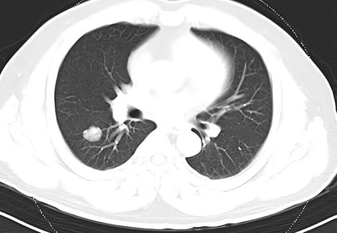
Routine follow-up in March 2022 revealed an incidental lung lesion on chest X-ray. CT (Computed Tomography) Scan, as shown in Fig. 1, and Positron Emission Tomography (PET) detected a 2.7 × 2.4 cm lesion in the right lower lobe of the lung (Maximum Standardized Uptake Value (SUV max)—17.04) and an enlarged right hilar node measuring 2.1 × 1.6 cm (SUV max 23.16). The possibilities considered included metastasis and a second primary. The PET-positive hilar lymph nodes with no other significant nodes suggested a possible second primary. Guided biopsy from the right lung mass showed metastatic adenocarcinoma with clear cell morphology. Immunohistochemistry (IHC) revealed diffuse strong positivity for cytokeratin (CK) and Paired-Box Gene 8 (PAX8), focal weak to moderate positivity for Cluster of Differentiation 10 (CD10), and negativity for CK7, supporting a diagnosis of metastatic clear cell RCC rather than primary lung adenocarcinoma [4].
Figure 1 CT Scan of the chest shows a minimally enhancing lesion in the right lower lobe lung.
The patient underwent right lower lobectomy and hilar lymph node dissection in May 2022 following cardiopulmonary evaluation and informed consent. Intraoperatively, a 2 × 3 cm lesion was observed in the right lower lobe and enlarged nodes with extranodal extension were found in 10R and 11R stations. Postoperative recovery was uneventful. Final histopathology confirmed metastatic renal cell carcinoma. Notably, 2 out of 4 excised nodes contained metastatic RCC. Detailed histopathology with IHC confirmed the origin of the metastases, excluding primary lung cancer involvement.
The patient was monitored regularly with a 2-year disease-free interval before diagnosis of multiple skeletal metastases, for which treatment is ongoing.
Case 2
A 47-year-old lady was evaluated in December 2020 elsewhere for early satiety, upper abdominal discomfort, and melena. Workup including endoscopy, biopsy, and contrast-enhanced CT revealed carcinoma of the stomach. She underwent a total gastrectomy with D2 lymphadenectomy. Histopathology showed poorly cohesive adenocarcinoma with signet ring cells infiltrating the serosa. All 16 lymph nodes were positive. Stage was pT4aN3bM0 (Stage IIIC). She completed adjuvant chemotherapy with oxaliplatin and capecitabine (CAPEOX) by August 2021 and was followed regularly.
In March 2022, a palpable left breast lump (~4 cm) with nipple retraction and axillary lymphadenopathy was noted. Mammogram classified the lesion as BIRADS 4b (Breast Imaging-Reporting and Data System) with significant lymph nodes. Differential diagnosis included second primary breast carcinoma versus rare breast metastasis. Core needle biopsy revealed poorly differentiated carcinoma with signet ring cells. IHC showed strong membranous positivity for CK, weak nuclear positivity for GATA binding protein 3 (GATA3), and was negative for estrogen receptor (ER), progesterone receptor (PR), and human epidermal growth factor receptor 2 (HER2), and CK20. Combined with the patient’s history, this favored metastatic adenocarcinoma with signet ring morphology from the gastric primary, ruling out primary breast carcinoma [5, 6, 7].
Treatment options of second-line systemic therapy versus metastasectomy were discussed at the tumour board. Given the short disease-free interval, second-line chemotherapy was initiated. The patient received six cycles of ramucirumab and paclitaxel till October 2022. Subsequent 18F-Fluorodeoxyglucose PET/CT showed no metabolically active disease despite persistent clinical breast lump. Metastasectomy was then offered.
Left modified radical mastectomy was performed in February 2023 due to the tumour involving the entire breast. Axillary dissection showed three of seventeen lymph nodes positive for metastases. Such nodal involvement from breast metastasis of gastric cancer is exceedingly rare.
Discussion
This case report aims to highlight the possibility of lymph node involvement in regional nodal basins of metastatic foci and offers several points for discussion: the mechanisms of hilar and axillary lymph node involvement in renal cell carcinoma (RCC) and gastric cancer, respectively; the incidence of such lymph node metastasis; the prognostic significance; and the role of surgery in the management of metastases and lymph nodes in these scenarios [3, 4].
A key question in this context is the mechanism of lymph node metastasis (LNM) occurring alongside metastatic foci, but in the absence of disease in other lymph node basins. The most plausible explanation is the concept of “re-metastasis”, which refers to locoregional spread of metastatic tumor cells from an existing metastatic site [3, 4]. For example, in the first case, hilar lymph node involvement likely represents lymphatic spread from the lung metastasis. Another, albeit rarer, explanation is that circulating tumour cells (CTCs) seed the lymph nodes in addition to the lung metastasis. Both of these explanations align with the fundamental models of metastasis—the linear progression model and the parallel progression model [2, 3].
According to these models, this unusual route of metastasis from a metastatic focus can be enumerated in both cases. Hilar lymph node involvement in lung metastasis from RCC is believed to arise from the metastatic focus itself, as there are no other nodal basins involved in the spread from the primary tumour. Similarly, axillary lymph node involvement in gastric adenocarcinoma may be explained through the same principle.
The diagnosis of breast metastasis from gastric signet ring cell carcinoma is a rare clinical entity posing diagnostic and therapeutic challenges. In this case, the breast lesion and axillary lymph node involvement were confirmed as metastatic adenocarcinoma of gastric origin, based on histopathology and immunohistochemistry. Although 3 of 17 axillary nodes were positive, raising the possibility of secondary spread from breast metastasis, it is critical to distinguish this, as it impacts staging, treatment, and prognosis significantly. Comprehensive immunophenotypic profiling, which included negative expression of breast markers such as ER, PR, HER2, and GATA3 alongside gastric cancer markers, helped exclude both primary breast carcinoma and secondary nodal spread from it [5, 6]. Such metastatic patterns emphasize the complexity of these cases and highlight the need for high clinical suspicion and thorough pathological evaluation to ensure accurate diagnosis.
Another important issue is the prognostic implication of metastatic lymph nodes from metastatic foci and the role of lymphadenectomy along with metastasectomy. Lymphadenectomy is not routinely performed with metastasectomy, making it difficult to determine the incidence and prognostic value of lymph node metastases in such contexts from existing literature. Data on breast metastasis from gastric signet ring cell carcinoma are limited, with only around 60 cases reported worldwide [5, 6, 8].
In lung metastasis from RCC, there is ongoing debate whether mediastinal lymphadenectomy should be performed during renal cell carcinoma lung metastasectomy. Existing evidence is derived from studies where lymph nodes were dissected based on preoperative imaging findings of enlarged or metabolically active nodes, or were inadvertently removed during metastasectomy [4, 9]. A summary of relevant data from these studies is provided in Table 1 (Ref. [9, 10, 11, 12, 13]), adapted from Stephane Renaud, 2013 [4].
Table 1: Incidence of mediastinal nodes in various series where mediastinal lymphadenectomy was done as part of pulmonary metastasectomy for Renal cell carcinoma.
| Authors | No. of Patients underwent LN Dissection | Incidence of lymph node metastasis | Outcome |
| Murthy et al. [10] (2004) | 32/92 (34.78%) | 12/32 (37.50%) | OS: significantly lower in case of LNI (65% in the absence of LNI vs. 30% when three nodes were involved, with a median follow-up of 3.7 years) |
| Pfannschmidt et al. [11] (2002) | 191/248 (77.00%) | 57/191 (29.84%) | Three-year OS: 31.4% (N+) vs. 55.4% (N0) LNI: independent prognostic factor (p = 0.0038) |
| Assouad et al. [12] (2007) | 44/65 (67.69%) | 13/44 (29.54%) | 5-year OS: pN+: 0% vs. pN−: 52% LNI: independent prognostic factor (p = 0.0018) |
| Winter et al. [9] (2010) | 110/156 (70.50%) | 38/110 (34.54%) | Mean survival of pN0: 102.2 months, pN+: 19.1 months (p < 0.001) Better OS in case of lymphadenectomy (p = 0.08) |
| Meimarakis et al. [13] (2010) | 91/202 (45.04%) | 27/91 (29.67%) | pN+: 19.1 (95% CI: 5.8–32.4) months vs. pN0: 92.0 (95% CI: 35.7–148.2) months; (p < 0.001) LNI: independent prognostic factor (p < 0.004) |
No.: Number; LN: Lymph Node; LNI: Lymph Node Involvement; OS: Overall Survival; N+: Number of positive lymph nodes; N0: Number of negative lymph nodes; pN+: Pathologically positive lymph nodes; pN− or pN0: Pathologically negative lymph nodes; CI: Confidence Interval.
In most of the aforementioned studies, lymph node involvement (LNI) was detected in approximately 30% of cases where lymphadenectomy was performed based on radiological suspicion [4]. Therefore, omitting lymphadenectomy risks failure to achieve complete resection and increases the likelihood of loco-regional recurrence. These studies consistently conclude that LNI is a significant independent prognostic factor for survival [4, 9].
Despite the poor prognosis associated with LNI, surgery remains the best potentially curative treatment option for RCC with metastases. Consequently, a systematic total mediastinal lymphadenectomy is recommended, even if preoperative imaging does not suggest lymph node invasion [9]. This approach is likely to improve loco-regional control and provide essential staging and prognostic information [14, 15]. However, current literature is limited and primarily based on low-level evidence, warranting further studies to confirm these recommendations.
Regarding our second case, although breast cancer incidence worldwide is approximately 2.1 million per year, metastasis to the breast is unusual. Only 0.5 to 2% of patients present with breast metastases, primarily originating from melanoma, lymphoma, lung, and ovarian cancers, with gastrointestinal tumours being among the least common sources [5, 16]. While systemic treatment is considered the primary intervention, its role remains controversial [17]. Prognosis for patients with breast metastases has improved recently due to advances in anticancer therapies. Conversely, available data suggest that surgery plays an unclear role, may not significantly improve survival, and is generally considered palliative. These patients face a poor prognosis, with mortality rates exceeding 80% within the first year [6, 8, 18]. Further research is necessary to elucidate the role of surgery, including lymphadenectomy, in managing such cases.
Several inherent limitations must be considered when interpreting this report and the accompanying review of the literature. First, the incredibly low global incidence of these specific metastatic pathways—such as breast metastasis from gastric signet ring cell carcinoma—restricts the available data to isolated case reports and small, retrospective series [5, 7]. This introduces significant publication bias, as unusual presentations with unique surgical outcomes are more likely to be documented than standard presentations. Second, the current medical literature lacks standardized, prospective clinical trials evaluating the survival benefit of systematic lymphadenectomy during metastasectomy in these rare cohorts. Much of the analyzed data regarding RCC lung metastasectomy relies on low-level evidence where lymph node dissections were not uniformly performed, introducing heavy selection bias based on varied preoperative imaging and individual surgeon preferences [4, 9].
Conclusion
These cases highlight the rare but significant phenomenon of secondary metastasis, where metastatic lesions themselves spread to regional lymph nodes. Recognizing this metastatic pattern is important for accurate staging, prognosis, and treatment planning. Careful pathological and immunohistochemical evaluation is essential to distinguish metastasis-from-metastasis versus primary or initial metastatic disease, influencing management strategies.
In renal cell carcinoma, lymph node metastasis during lung metastasectomy is an adverse prognostic factor, supporting consideration of systematic lymphadenectomy for better local control and staging. However, in the case of breast metastasis from gastric cancer, surgery remains mainly palliative with limited survival benefit, emphasizing the need for individualized multidisciplinary care.
Overall, awareness of re-metastasis pathways and thorough clinical evaluation can help optimize oncologic outcomes in complex metastatic scenarios. Further research is needed to clarify the therapeutic role of lymphadenectomy in these uncommon presentations.
Availability of data and materials
The data supporting the findings of this study are included within the article. Further details or additional datasets are available from the corresponding author upon reasonable request.
Author contributions
MS—Conceptualization; Data curation; Formal analysis; Writing–original draft; Writing–review & editing. VG—Conceptualization; Data acquisition; Investigation; Writing–review & editing. MM—Conceptualization; Data curation; Validation; Writing–review & editing; Supervision. All authors read and approved the final manuscript and agree to be accountable for all aspects of the work.
Ethics approval and consent to participate
The permission was taken from the institutional ethics committee (Regional Cancer Centre, Thiruvananthapuram, Kerala) before starting the project. All procedures performed in studies involving human participants were in accordance with the ethical standards of the institutional research Board (Regional Cancer Centre, Thiruvananthapuram, Kerala) and with the 1964 Helsinki Declaration and its later amendments or comparable ethical standards. Informed consent was obtained from both participants included in the study.
Acknowledgment
Not applicable.
Funding
This research received no external funding.
Conflict of interest
The authors declare no conflict of interest.
REFERENCES
1. Welch DR. Defining a cancer metastasis. In Teicher BA (ed.) AACR Education Book (pp. 111–115). American Association for Cancer Research: Philadelphia. 2006.
2. Welch DR, Hurst DR. Defining the hallmarks of metastasis. Cancer Research. 2019; 79: 3011–3027.
3. Koscielny S, Tubiana M. Parallel progression of tumour and metastases. Nature Reviews Cancer. 2010; 10: 156.
4. Renaud S, Falcoz PE, Olland A, Massard G. Should mediastinal lymphadenectomy be performed during lung metastasectomy of renal cell carcinoma? Interactive CardioVascular and Thoracic Surgery. 2013; 16: 525–528.
5. Iesato A, Oba T, Ono M, Hanamura T, Watanabe T, Ito T, et al. Breast metastases of gastric signet-ring cell carcinoma: a report of two cases and review of the literature. OncoTargets and Therapy. 2014; 8: 91–97.
6. Lordick F, Carneiro F, Cascinu S, Fleitas T, Haustermans K, Piessen G, et al. Gastric cancer: ESMO clinical practice guideline for diagnosis, treatment and follow-up. Annals of Oncology. 2022; 33: 1005–1020.
7. Ding J, Gu H, Yang Z, Lu Y, Guo G. Breast metastasis from lung adenocarcinoma: a case report and review of the literature. Frontiers in Oncology. 2024; 14: 1370453.
8. Ma Y, Liu W, Li J, Xu Y, Wang H. Gastric cancer with breast metastasis: clinical features and prognostic factors. Oncology Letters. 2018; 16: 5565–5574.
9. Winter H, Meimarakis G, Angele MK, Hummel M, Staehler M, Hoffmann RT, et al. Tumor infiltrated hilar and mediastinal lymph nodes are an independent prognostic factor for decreased survival after pulmonary metastasectomy in patients with renal cell carcinoma. Journal of Urology. 2010; 184: 1888–1894.
10. Murthy SC, Kim K, Rice TW, Rajeswaran J, Bukowski R, DeCamp MM, et al. Can we predict long-term survival after pulmonary metastasectomy for renal cell carcinoma? The Annals of Thoracic Surgery. 2005; 79: 996–1003.
11. Pfannschmidt J, Hoffmann H, Muley T, Krysa S, Trainer C, Dienemann H. Prognostic factors for survival after pulmonary resection of metastatic renal cell carcinoma. The Annals of Thoracic Surgery. 2002; 74: 1653–1657.
12. Assouad J, Petkova B, Berna P, Dujon A, Foucault C, Riquet M. Renal cell carcinoma lung metastases surgery: pathologic findings and prognostic factors. The Annals of Thoracic Surgery. 2007; 84: 1114–1120.
13. Meimarakis G, Angele M, Staehler M, Clevert DA, Crispin A, Rüttinger D, et al. Evaluation of a new prognostic score (Munich score) to predict long-term survival after resection of pulmonary renal cell carcinoma metastases. American Journal of Surgery. 2011; 202: 158–167.
14. National Cancer Institute. Renal cell cancer treatment (PDQ®)–health professional version. 2025. Available at: https://www.cancer.gov/types/kidney/hp/kidney-treatment-pdq (Accessed: 21 October 2025).
15. Bendimya M, Kairouani M, El Magroud M, Bennani A, Al Jarroudi O, Brahmi SA, et al. Unusual metastasis of gastric signet ring cell carcinoma to the breast: a case report of a young Moroccan patient. Cureus. 2024; 16: e56333.
16. Khalayleh H, Bader R, Imam A, Khoury D, Yosepovich A, Bar-Zakai B, et al. Gastric cancer metastases to the breast: systemic review and meta-analysis of the English literature. Chirurgia. 2023; 36: 261–269.
17. Buerba-Vieregge HH, Fernández-Ferreira R, Soberanis-Piña PD, De la Peña-López IR, Navarro-García LM, Macari-Jorge A. Breast metastasis of gastric signet ring cell carcinoma: a case report and literature review. Case Reports in Oncology. 2021; 14: 165–172.
18. Ajani JA, D’Amico TA, Bentrem DJ, Chao J, Cooke D, Corvera C, et al. Gastric cancer, version 2.2022, NCCN clinical practice guidelines in oncology. Journal of the National Comprehensive Cancer Network. 2022; 20: 167–192.